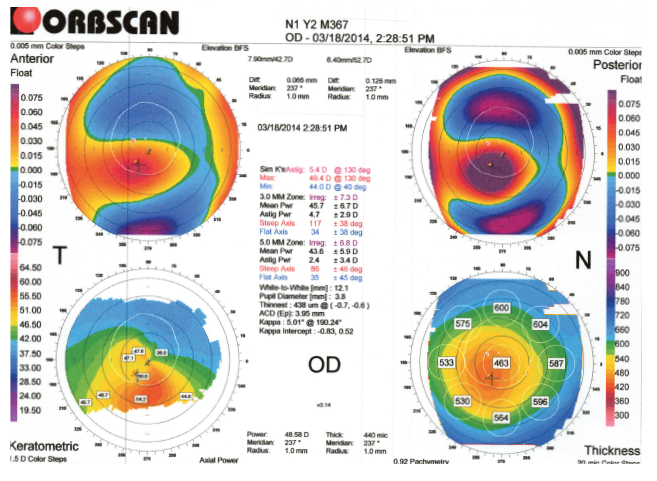

The numerical map shows greater than 3.00D of inferior/superior discrepancy. Topography revealed inferior steepening that at first glance resembles an ectasia-like pattern in the left eye (Figure 1). The left eye refracted to -2.75 +1.75 X 090 for 20/25 BSCVA acuity. The right eye revealed a refraction of -4.00 +0.50 X 085 for 20/15 BSCVA. In this article, I want to elucidate several nuances regarding the diagnosis of keratoconus/forme fruste keratoconus and false-positive diagnoses for both entities.Ī 38-year-old white male presented with an irregular astigmatism pattern in his left eye. As physicians and surgeons, we do not treat patients based solely upon a printout. Even then, one is only making a diagnosis from a piece of paper. I cannot however, I agree that most experienced ophthalmologists can diagnose keratoconus, forme fruste keratoconus, pellucid marginal degeneration, or keratectasia-like “patterns” from a quarter-mile away with a pair of binoculars.

Some ophthalmologists say they can make the diagnosis of keratoconus, forme fruste keratoconus, pellucid marginal degeneration, or keratectasia by topography analysis alone. Refractive IOLs: Are You and Your ASC Ready? Trachoma: Still A Major Cause of Preventable Blindness Reflective Particles in the Cataract IncisionĬhildhood Blindness and Pediatric Cataract Transitioning to Bimanual Microincisional Phacoemulsification

Peer-Reviewed Literature: The Femtosecond Laser


 0 kommentar(er)
